Moles and skin lesions come in variety of shapes and sizes and cause a variety of problems. Often the most common reason for removal is that the client does not like the way the mole, cyst or other skin lesion looks and this causes them to feel self-conscious or embarrassed. Other reasons include a change in the appearance of the mole that makes them concerned it may be a skin cancer. Sometimes the client doesn’t notice the change in the mole but their partner, relative or friend notices and brings the change in the appearance of the mole to their attention. The system used to see whether moles may be a skin cancer is called the ABCD of moles.
A stands for Asymmetry. If the mole had an imaginary vertical line drawn through the middle of it, then both halves of the mole should look the same shape. If either half of the mole looks different to the other half then further investigation may be required.
B stands for border irregularity. The border of a mole is normally smooth, but if it is notched then it could mean the mole has changed into something that needs attention such as a skin cancer.
C stands for colour irregularity. A mole should be the same shade and colour throughout. If the mole has different shades of pigment then it could mean the mole has changed and needs investigating.
D stands for increasing diameter i.e. the mole is increasing in size. This usually refers to the horizontal measurement although moles can also grow vertically and increase in height forming a nodule.
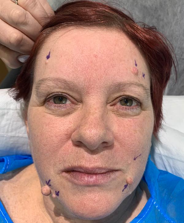
Safe Mole Removal
The aim of mole removal is to provide a medically safe removal of the mole that is appropriate for the character of mole and also provide an optimal scar. I would recommend histology for any mole or skin lesion removal so the diagnosis can be confirmed. However, some clients decline this and this is at their own personal risk.
Types of Procedure
Histology
Although the majority of moles that are examined appear to be non-cancerous, occasionally, a mole can be removed that was thought to be non-cancerous but is actually a skin cancer. The only way to know for sure is to have the mole analysed. It is the client’s choice whether they would prefer to have the mole analysed as it is an additional charge but that is the client’s own decision and personal risk. Histology provides microscopic evidence to support the clinical diagnosis made on the history and examination during the consultation.
Your Outpatient Visit
This procedure is performed as outpatient in a high-quality state of the art operating theatre with a trained nurse present.
Typical Duration Of Surgery
A single mole may take about 15 minutes to remove. The more moles that need removing, the longer the time it takes.
Anaesthesia Required
Skin lesions are removed by numbing the skin with a small injection. This is called local anaesthetic.
Healing Time
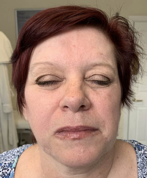
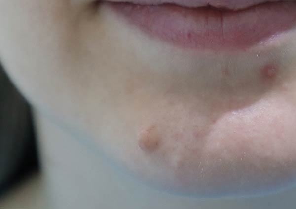
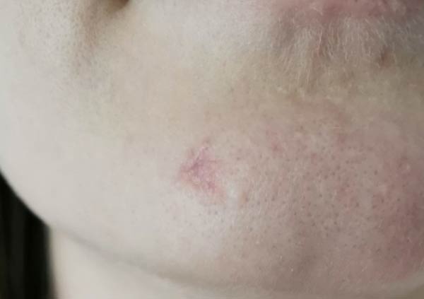
The wounds have stitches that need removing at about a week. Clients can shower as normal the evening of surgery as there is a shower-proof dressing over the wound. After two weeks, scars should be massaged (to help flatten the scar), moisturised (to prevent the scar becoming dry) and sunblock applied over the scar (to prevent the scar from tanning differently).
Risks associated with the procedure
- Bleeding (haematoma). Occasionally, after surgery, blood may pool under the skin (haematoma), this usually disperses spontaneously over 2 to 3 weeks, but very rarely needs to be drained surgically if larger and fails to disperse.
- It is important to recognise that a surgical incision contributes to a risk of the introduction of bacteria from the patient’s own skin. Such infection may be associated with tiredness, weakness, fever and muscle aches and pains. Antibiotics are not normally given during this procedure and there is no evidence that by giving antibiotics this reduces the chance of an infection occurring. If you experience any symptoms of infection, contact your clinic nurse or the advised out of hours contact number.
- Wound healing. Healing of wounds varies from patient to patient and even from one part of the body to another and is a gradual process. Smoking for the period leading up to your surgery and afterwards can seriously hinder the healing process.
- Small cysts called milia can form along the scar line. This is normally temporary, but sometimes may need surgical removal
- Scar variability and keloids. All prospective patients must be aware that the rate at which their scars may heal and fade are entirely variable and individual. Uncommonly, a scar does not heal in the normal way. This is known as hypertrophic scarring meaning the scar is excessively thick. Scars may be red, or highly coloured, thick, painful and may take several years to improve if at all. Although this an unusual problem, it cannot be avoided or diagnosed in advance.
- Incomplete removal. Moles may not be completely removed. As stated, a shave biopsy does not completely remove the mole and there is high chance of the mole appearing again. Sometimes a mole may look like it has been removed using the naked eye, but when the mole is analysed under the microscope, it is found that either the outer edge of the mole or base of the mole is still there. This is why histology is an important tool is confirming complete removal.
- Diagnosis of a skin cancer. It is rare that a mole that looks completely harmless according to the ABCD system is a skin cancer. However, sometimes, a mole is sent to histology and comes back confirming a form of skin cancer. If this occurs, a discussion will take place on the best course of management.
Cysts are common and are fluid filled sacs within or under the skin. They can become red, infected and cause pain as well as making a client self-conscious about their appearance. The commonest types of cyst are epidermal cysts and these are commonly found on the face and back. They occur when the hair follicles get blocked and the oils that would normally be secreted along the hair follicle and skin accumulate in their sac of origin causing the sac to swell.